Dental Operating Microscope

Dental Treatment with a Dental Operating Microscope: Perfect Vision, Maximum Protection

Dental operating microscope-assisted treatments, representing one of the most advanced technological advancements in dentistry, offer a whole new dimension by surpassing the limitations of the naked eye. Thanks to advanced dental operating microscopes that can magnify the working field 6, 15, or even 40 times, the margin of error in diagnosis and treatment is virtually eliminated. This enormous magnification capacity, combined with a powerful light source, allows the dentist to "see exactly what they are doing in the finest detail" at every stage, from the most complex root canals to millimeter-precise aesthetic touches.

The Transition from Traditional to Modern: Why a Dental Operating Microscope?

The "Extension For Prevention" approach, which dominated dentistry in the past, has now been replaced by a "Minimally Invasive" approach (with minimal tissue loss) thanks to dental operating microscope technology. Previously, dentists had to remove extra tissue from the tooth just to see the decay, be sure, and ensure the retention of old filling materials. Today, thanks to the 40x magnification provided by dental operating microscopes, precise cleaning can be performed by targeting only the diseased area without touching healthy tissue.

As David Clark, founder of the Academy of Microscope-Assisted Dentistry, stated:

“The greatest indicator of the long-term retention of teeth in the mouth is the volume of healthy and natural tooth tissue remaining after treatment.” Endodontics academies such as AAE and ESE recommend the use of a Dental Operating Microscope in routine cases and make it mandatory in specialist training. At Dental Esthetic Center, with the help of a dental operating microscope, we remove only as much tissue as necessary from your tooth, maximizing the lifespan of our restorations.

The Unique Advantages of Dental Surgical Microscopes in Treatment

Dental operating microscopes are used in our clinic in many areas, from routine procedures to the most challenging cases:

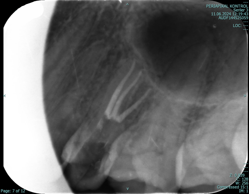
• Discovering the Hidden Details in Root Canal Treatments: Root canals are extremely complex and capillary structures. Thanks to a dental microscope, extra canals, narrowed or blocked structures that are impossible to see with the naked eye are detected instantly. As a result, the success rate of the first root canal treatment is much higher compared to traditional methods.

• A Critical Role in Root Canal Restoration (Retreatment) Therapies: Retreating (removing and redoing) previously performed but failed root canal treatments that have developed infection is one of the most challenging procedures in dentistry. A dental operating microscope is indispensable for the complete removal of old root canal fillings, the safe removal of any broken instruments within the canal, and the identification and treatment of any missed extra canals that may have caused the failure.

• Precise and Early Detection of Tooth Cracks: Micro-cracks, undetectable to the naked eye or even with standard X-rays, are often the root cause of unexplained toothaches (especially during chewing). A dental microscope allows us to precisely determine the size, direction, and depth of these hairline cracks. This enables early diagnosis before the crack progresses and the tooth needs extraction, allowing for the application of the correct treatment protocol and saving the tooth.

• Flawless Precision and Aesthetics: In porcelain and composite veneer applications, the precise and smooth cuts (steps) made on the tooth are adjusted under a dental operating microscope. This guarantees much more natural aesthetic results and a perfect fit of the restoration to the tooth. • Extensive Application Area: It provides unparalleled precision not only in root canal treatment or caries cleaning, but also in gum treatments, checking the accuracy of impressions, tartar removal, and delicate operations.

How does our dental microscope-assisted treatment process progress? 

  1. Detailed Review: The source of your complaint (cracks, hidden cavities, root canal problems) is examined in detail using a dental operating microscope under high magnification and powerful lighting.
  2. Precise Treatment Planning: Based on this enhanced data, where no detail is overlooked, the safest and most protective treatment plan is created.
  3. Minimally Invasive ProcedureThe procedure is performed with high precision only on the target area, causing minimal disturbance to surrounding tissues.
  4. Secure Tracking: Post-treatment compliance and success are monitored with the same care to ensure long-lasting results.


In summaryTreating small and delicate teeth requires an equally high level of precision. The more detailed a dentist's field of vision, the more flawless the treatments can be. With our dental microscope-assisted approach, we save teeth that were previously deemed "necessary to be extracted," ensuring your teeth remain healthy in your mouth for many years to come. Because for us, clearer vision means more perfect treatment.

Contact Us

We want to create a positive and comfortable environment for you in our clinic during your stay and ensure a healthy and trouble-free treatment process.

Visit Us

Oran Mahallesi Kudüs Caddesi Park Oran Ofis 1/21 Kat:16 Daire No:54 Çankaya / ANKARA

Our Working Hours

Monday – Friday

09:30 - 18:30

Saturday

09:00 - 15:30

Sunday

Closed